Misplaced Pages

40S ribosomal protein S27a

Article snapshot taken from Wikipedia with creative commons attribution-sharealike license. Give it a read and then ask your questions in the chat. We can research this topic together.
(Redirected from RPS27A) Protein-coding gene in the species Homo sapiens

RPS27A
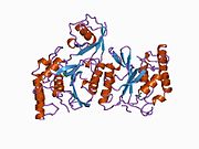
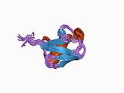
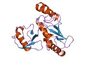
Available structures
PDBOrtholog search: PDBe RCSB
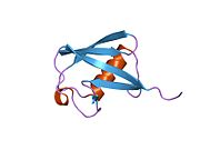
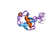
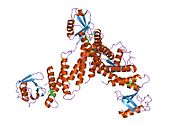
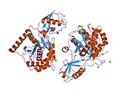
List of PDB id codes

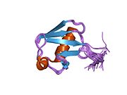
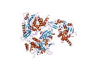
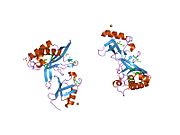
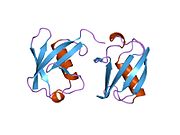
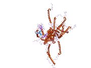
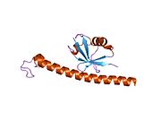
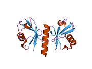
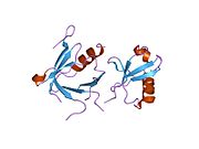
2KTF, 2L0F, 2L0T, 2XK5, 3AXC, 3NHE, 3NOB, 3PHW, 3TBL, 4UG0, 4V6X, 5AJ0, 4R62, 5FLX, 4UJD, 4KZZ, 4UJC, 3J7P, 4KZY, 4D5L, 3J7R, 4D61, 4KZX, 4UJE, 5A2Q

Identifiers
AliasesRPS27A, CEP80, HEL112, S27A, UBA80, UBC, UBCEP1, UBCEP80, ribosomal protein S27a
External IDsOMIM: 191343; MGI: 1925544; HomoloGene: 37715; GeneCards: RPS27A; OMA:RPS27A - orthologs
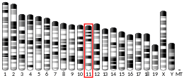
Gene location (Human)
Chromosome 2 (human)
Chr.Chromosome 2 (human)
Chromosome 2 (human)Genomic location for RPS27AGenomic location for RPS27A
Band2p16.1Start55,231,903 bp
End55,235,853 bp
Gene location (Mouse)
Chromosome 11 (mouse)
Chr.Chromosome 11 (mouse)
Chromosome 11 (mouse)Genomic location for RPS27AGenomic location for RPS27A
Band11|11 A3.3Start29,495,846 bp
End29,498,109 bp
RNA expression pattern
Bgee
HumanMouse (ortholog)
Top expressed in
  • left ovary

  • granulocyte

  • ganglionic eminence

  • right uterine tube

  • canal of the cervix

  • right ovary

  • body of uterus

  • ventricular zone

  • left lobe of thyroid gland

  • anterior pituitary
Top expressed in
  • embryo

  • embryo

  • epiblast

  • urinary bladder

  • bone marrow

  • ventricular zone

  • uterus

  • pancreas

  • adrenal gland

  • lens
More reference expression data
BioGPS
n/a
Gene ontology
Molecular function
Cellular component
Biological process
Sources:Amigo / QuickGO
Orthologs
SpeciesHumanMouse
Entrez

6233

78294

Ensembl

ENSG00000143947

ENSMUSG00000020460

UniProt

P62979

P62983

RefSeq (mRNA)

NM_002954
NM_001135592
NM_001177413

NM_001033865
NM_024277

RefSeq (protein)

NP_001129064
NP_001170884
NP_002945

NP_001029037
NP_077239

Location (UCSC)Chr 2: 55.23 – 55.24 MbChr 11: 29.5 – 29.5 Mb
PubMed search
Wikidata
View/Edit HumanView/Edit Mouse

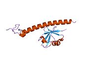
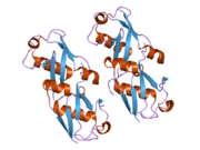
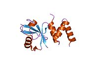
40S ribosomal protein S27a is a protein that in humans is encoded by the RPS27A gene.

Ubiquitin, a highly conserved protein that has a major role in targeting cellular proteins for degradation by the 26S proteosome, is synthesized as a precursor protein consisting of either polyubiquitin chains or a single ubiquitin fused to an unrelated protein. This gene encodes a fusion protein consisting of ubiquitin at the N terminus and ribosomal protein S27a at the C terminus. When expressed in yeast, the protein is post-translationally processed, generating free ubiquitin monomer and ribosomal protein S27a. Ribosomal protein S27a is a component of the 40S subunit of the ribosome and belongs to the S27AE family of ribosomal proteins. It contains C4-type zinc finger domains and is located in the cytoplasm. Pseudogenes derived from this gene are present in the genome. As with ribosomal protein S27a, ribosomal protein L40 is also synthesized as a fusion protein with ubiquitin; similarly, ribosomal protein S30 is synthesized as a fusion protein with the ubiquitin-like protein fubi.

References

  1. ^ GRCh38: Ensembl release 89: ENSG00000143947Ensembl, May 2017
  2. ^ GRCm38: Ensembl release 89: ENSMUSG00000020460Ensembl, May 2017
  3. "Human PubMed Reference:". National Center for Biotechnology Information, U.S. National Library of Medicine.
  4. "Mouse PubMed Reference:". National Center for Biotechnology Information, U.S. National Library of Medicine.
  5. Kenmochi N, Kawaguchi T, Rozen S, Davis E, Goodman N, Hudson TJ, Tanaka T, Page DC (Aug 1998). "A map of 75 human ribosomal protein genes". Genome Res. 8 (5): 509–23. doi:10.1101/gr.8.5.509. PMID 9582194.
  6. ^ "Entrez Gene: RPS27A ribosomal protein S27a".

Further reading

PDB gallery
  • 1aar: STRUCTURE OF A DIUBIQUITIN CONJUGATE AND A MODEL FOR INTERACTION WITH UBIQUITIN CONJUGATING ENZYME (E2) 1aar: STRUCTURE OF A DIUBIQUITIN CONJUGATE AND A MODEL FOR INTERACTION WITH UBIQUITIN CONJUGATING ENZYME (E2)
  • 1cmx: STRUCTURAL BASIS FOR THE SPECIFICITY OF UBIQUITIN C-TERMINAL HYDROLASES 1cmx: STRUCTURAL BASIS FOR THE SPECIFICITY OF UBIQUITIN C-TERMINAL HYDROLASES
  • 1d3z: UBIQUITIN NMR STRUCTURE 1d3z: UBIQUITIN NMR STRUCTURE
  • 1f9j: STRUCTURE OF A NEW CRYSTAL FORM OF TETRAUBIQUITIN 1f9j: STRUCTURE OF A NEW CRYSTAL FORM OF TETRAUBIQUITIN
  • 1fxt: STRUCTURE OF A CONJUGATING ENZYME-UBIQUITIN THIOLESTER COMPLEX 1fxt: STRUCTURE OF A CONJUGATING ENZYME-UBIQUITIN THIOLESTER COMPLEX
  • 1g6j: STRUCTURE OF RECOMBINANT HUMAN UBIQUITIN IN AOT REVERSE MICELLES 1g6j: STRUCTURE OF RECOMBINANT HUMAN UBIQUITIN IN AOT REVERSE MICELLES
  • 1gjz: SOLUTION STRUCTURE OF A DIMERIC N-TERMINAL FRAGMENT OF HUMAN UBIQUITIN 1gjz: SOLUTION STRUCTURE OF A DIMERIC N-TERMINAL FRAGMENT OF HUMAN UBIQUITIN
  • 1nbf: Crystal structure of a UBP-family deubiquitinating enzyme in isolation and in complex with ubiquitin aldehyde 1nbf: Crystal structure of a UBP-family deubiquitinating enzyme in isolation and in complex with ubiquitin aldehyde
  • 1ogw: SYNTHETIC UBIQUITIN WITH FLUORO-LEU AT 50 AND 67 1ogw: SYNTHETIC UBIQUITIN WITH FLUORO-LEU AT 50 AND 67
  • 1otr: Solution Structure of a CUE-Ubiquitin Complex 1otr: Solution Structure of a CUE-Ubiquitin Complex
  • 1p3q: Mechanism of Ubiquitin Recognition by the CUE Domain of VPS9 1p3q: Mechanism of Ubiquitin Recognition by the CUE Domain of VPS9
  • 1q0w: Solution structure of Vps27 amino-terminal UIM-ubiquitin complex 1q0w: Solution structure of Vps27 amino-terminal UIM-ubiquitin complex
  • 1q5w: Ubiquitin Recognition by Npl4 Zinc-Fingers 1q5w: Ubiquitin Recognition by Npl4 Zinc-Fingers
  • 1s1q: TSG101(UEV) domain in complex with Ubiquitin 1s1q: TSG101(UEV) domain in complex with Ubiquitin
  • 1sif: Crystal structure of a multiple hydrophobic core mutant of ubiquitin 1sif: Crystal structure of a multiple hydrophobic core mutant of ubiquitin
  • 1tbe: STRUCTURE OF TETRAUBIQUITIN SHOWS HOW MULTIUBIQUITIN CHAINS CAN BE FORMED 1tbe: STRUCTURE OF TETRAUBIQUITIN SHOWS HOW MULTIUBIQUITIN CHAINS CAN BE FORMED
  • 1ubi: SYNTHETIC STRUCTURAL AND BIOLOGICAL STUDIES OF THE UBIQUITIN SYSTEM. PART 1 1ubi: SYNTHETIC STRUCTURAL AND BIOLOGICAL STUDIES OF THE UBIQUITIN SYSTEM. PART 1
  • 1ubq: STRUCTURE OF UBIQUITIN REFINED AT 1.8 ANGSTROMS RESOLUTION 1ubq: STRUCTURE OF UBIQUITIN REFINED AT 1.8 ANGSTROMS RESOLUTION
  • 1ud7: SOLUTION STRUCTURE OF THE DESIGNED HYDROPHOBIC CORE MUTANT OF UBIQUITIN, 1D7 1ud7: SOLUTION STRUCTURE OF THE DESIGNED HYDROPHOBIC CORE MUTANT OF UBIQUITIN, 1D7
  • 1uzx: A COMPLEX OF THE VPS23 UEV WITH UBIQUITIN 1uzx: A COMPLEX OF THE VPS23 UEV WITH UBIQUITIN
  • 1v80: Solution structures of ubiquitin at 30 bar and 3 kbar 1v80: Solution structures of ubiquitin at 30 bar and 3 kbar
  • 1v81: Solution structures of ubiquitin at 30 bar and 3 kbar 1v81: Solution structures of ubiquitin at 30 bar and 3 kbar
  • 1wr1: The complex structure of Dsk2p UBA with ubiquitin 1wr1: The complex structure of Dsk2p UBA with ubiquitin
  • 1wr6: Crystal structure of GGA3 GAT domain in complex with ubiquitin 1wr6: Crystal structure of GGA3 GAT domain in complex with ubiquitin
  • 1wrd: Crystal structure of Tom1 GAT domain in complex with ubiquitin 1wrd: Crystal structure of Tom1 GAT domain in complex with ubiquitin
  • 1xd3: Crystal structure of UCHL3-UbVME complex 1xd3: Crystal structure of UCHL3-UbVME complex
  • 1xqq: Simultaneous determination of protein structure and dynamics 1xqq: Simultaneous determination of protein structure and dynamics
  • 1yd8: COMPLEX OF HUMAN GGA3 GAT DOMAIN AND UBIQUITIN 1yd8: COMPLEX OF HUMAN GGA3 GAT DOMAIN AND UBIQUITIN
  • 1yiw: X-ray Crystal Structure of a Chemically Synthesized Ubiquitin 1yiw: X-ray Crystal Structure of a Chemically Synthesized Ubiquitin
  • 1yj1: X-ray Crystal Structure of a Chemically Synthesized Ubiquitin 1yj1: X-ray Crystal Structure of a Chemically Synthesized Ubiquitin
  • 1yx5: Solution Structure of S5a UIM-1/Ubiquitin Complex 1yx5: Solution Structure of S5a UIM-1/Ubiquitin Complex
  • 1yx6: Solution Structure of S5a UIM-2/Ubiquitin Complex 1yx6: Solution Structure of S5a UIM-2/Ubiquitin Complex
  • 1zgu: Solution structure of the human Mms2-Ubiquitin complex 1zgu: Solution structure of the human Mms2-Ubiquitin complex
  • 2ayo: Structure of USP14 bound to ubquitin aldehyde 2ayo: Structure of USP14 bound to ubquitin aldehyde
  • 2bgf: NMR STRUCTURE OF LYS48-LINKED DI-UBIQUITIN USING CHEMICAL SHIFT PERTURBATION DATA TOGETHER WITH RDCS AND 15N-RELAXATION DATA 2bgf: NMR STRUCTURE OF LYS48-LINKED DI-UBIQUITIN USING CHEMICAL SHIFT PERTURBATION DATA TOGETHER WITH RDCS AND 15N-RELAXATION DATA
  • 2c7m: HUMAN RABEX-5 RESIDUES 1-74 IN COMPLEX WITH UBIQUITIN 2c7m: HUMAN RABEX-5 RESIDUES 1-74 IN COMPLEX WITH UBIQUITIN
  • 2c7n: HUMAN RABEX-5 RESIDUES 1-74 IN COMPLEX WITH UBIQUITIN 2c7n: HUMAN RABEX-5 RESIDUES 1-74 IN COMPLEX WITH UBIQUITIN
  • 2d3g: Double sided ubiquitin binding of Hrs-UIM 2d3g: Double sided ubiquitin binding of Hrs-UIM
  • 2den: Solution Structure of the Ubiquitin-Associated Domain of Human BMSC-UbP and its Complex with Ubiquitin 2den: Solution Structure of the Ubiquitin-Associated Domain of Human BMSC-UbP and its Complex with Ubiquitin
  • 2dx5: The complex structure between the mouse EAP45-GLUE domain and ubiquitin 2dx5: The complex structure between the mouse EAP45-GLUE domain and ubiquitin
  • 2fcm: X-ray Crystal Structure of a Chemically Synthesized Ubiquitin with a Cubic Space Group 2fcm: X-ray Crystal Structure of a Chemically Synthesized Ubiquitin with a Cubic Space Group
  • 2fcn: X-ray Crystal Structure of a Chemically Synthesized Ubiquitin with a Cubic Space Group 2fcn: X-ray Crystal Structure of a Chemically Synthesized Ubiquitin with a Cubic Space Group
  • 2fcq: X-ray Crystal Structure of a Chemically Synthesized Ubiquitin with a Cubic Space Group 2fcq: X-ray Crystal Structure of a Chemically Synthesized Ubiquitin with a Cubic Space Group
  • 2fcs: X-ray Crystal Structure of a Chemically Synthesized Ubiquitin with a Cubic Space Group 2fcs: X-ray Crystal Structure of a Chemically Synthesized Ubiquitin with a Cubic Space Group
  • 2fid: Crystal Structure of a Bovine Rabex-5 fragment complexed with ubiquitin 2fid: Crystal Structure of a Bovine Rabex-5 fragment complexed with ubiquitin
  • 2fif: Crystal Structure of a Bovine Rabex-5 fragment complexed with ubiquitin 2fif: Crystal Structure of a Bovine Rabex-5 fragment complexed with ubiquitin
  • 2fuh: Solution Structure of the UbcH5c/Ub Non-covalent Complex 2fuh: Solution Structure of the UbcH5c/Ub Non-covalent Complex
  • 2g3q: Solution Structure of Ede1 UBA-ubiquitin complex 2g3q: Solution Structure of Ede1 UBA-ubiquitin complex
  • 2g45: Co-crystal structure of znf ubp domain from the deubiquitinating enzyme isopeptidase T (isot) in complex with ubiquitin 2g45: Co-crystal structure of znf ubp domain from the deubiquitinating enzyme isopeptidase T (isot) in complex with ubiquitin
  • 2gbj: Crystal Structure of the 9-10 8 Glycine Insertion Mutant of Ubiquitin. 2gbj: Crystal Structure of the 9-10 8 Glycine Insertion Mutant of Ubiquitin.
  • 2gbk: Crystal Structure of the 9-10 MoaD Insertion Mutant of Ubiquitin 2gbk: Crystal Structure of the 9-10 MoaD Insertion Mutant of Ubiquitin
  • 2gbm: Crystal Structure of the 35-36 8 Glycine Insertion Mutant of Ubiquitin 2gbm: Crystal Structure of the 35-36 8 Glycine Insertion Mutant of Ubiquitin
  • 2gbn: Crystal Structure of the 35-36 8 Glycine Insertion Mutant of Ubiquitin 2gbn: Crystal Structure of the 35-36 8 Glycine Insertion Mutant of Ubiquitin
  • 2gbr: Crystal Structure of the 35-36 MoaD Insertion Mutant of Ubiquitin 2gbr: Crystal Structure of the 35-36 MoaD Insertion Mutant of Ubiquitin
  • 2gmi: Mms2/Ubc13~Ubiquitin 2gmi: Mms2/Ubc13~Ubiquitin
  • 2hd5: USP2 in complex with ubiquitin 2hd5: USP2 in complex with ubiquitin
  • 2hth: Structural basis for ubiquitin recognition by the human EAP45/ESCRT-II GLUE domain 2hth: Structural basis for ubiquitin recognition by the human EAP45/ESCRT-II GLUE domain
  • 2ibi: Covalent Ubiquitin-USP2 Complex 2ibi: Covalent Ubiquitin-USP2 Complex
  • 2j7q: CRYSTAL STRUCTURE OF THE UBIQUITIN-SPECIFIC PROTEASE ENCODED BY MURINE CYTOMEGALOVIRUS TEGUMENT PROTEIN M48 IN COMPLEX WITH A UBQUITIN-BASED SUICIDE SUBSTRATE 2j7q: CRYSTAL STRUCTURE OF THE UBIQUITIN-SPECIFIC PROTEASE ENCODED BY MURINE CYTOMEGALOVIRUS TEGUMENT PROTEIN M48 IN COMPLEX WITH A UBQUITIN-BASED SUICIDE SUBSTRATE
  • 2nr2: The MUMO (minimal under-restraining minimal over-restraining) method for the determination of native states ensembles of proteins 2nr2: The MUMO (minimal under-restraining minimal over-restraining) method for the determination of native states ensembles of proteins
  • 2o6v: Crystal structure and solution NMR studies of Lys48-linked tetraubiquitin at neutral pH 2o6v: Crystal structure and solution NMR studies of Lys48-linked tetraubiquitin at neutral pH
  • 2oob: crystal structure of the UBA domain from Cbl-b ubiquitin ligase in complex with ubiquitin 2oob: crystal structure of the UBA domain from Cbl-b ubiquitin ligase in complex with ubiquitin
Protein biosynthesis: translation (bacterial, archaeal, eukaryotic)
Proteins
Initiation factor
Bacterial
Mitochondrial
Archaeal
Eukaryotic
eIF1
eIF2
eIF3
eIF4
eIF5
eIF6
Elongation factor
Bacterial/Mitochondrial
Archaeal/Eukaryotic
Release factor
Ribosomal Proteins
Cytoplasmic
60S subunit
40S subunit
Mitochondrial
39S subunit
28S subunit
Other concepts
Ribosomal RNA / ribosome subunits
Archaea
(70S)
Large (50S):
Small (30S):
Bacteria
(70S)
Large (50S):
Small (30S):
Eukaryotes
Cytoplasmic (80S)Large (60S):
Small (40S):
Mitochondrial (55S)Large (28S):
Small (39S):
Chloroplast (70S)Large (50S):
Small (30S):
Ribosomal proteins(See article table)
Stub icon

This article on a gene on human chromosome 2 is a stub. You can help Misplaced Pages by expanding it.

Stub icon

This protein-related article is a stub. You can help Misplaced Pages by expanding it.

Categories:
40S ribosomal protein S27a Add topic