| Ventral lateral nucleus | |
|---|---|
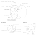
 Thalamic nuclei: Thalamic nuclei: MNG = Midline nuclear group AN = Anterior nuclear group MD = Medial dorsal nucleus VNG = Ventral nuclear group VA = Ventral anterior nucleus VL = Ventral lateral nucleus VPL = Ventral posterolateral nucleus VPM = Ventral posteromedial nucleus LNG = Lateral nuclear group PUL = Pulvinar MTh = Metathalamus LG = Lateral geniculate nucleus MG = Medial geniculate nucleus | |
 Thalamic nuclei Thalamic nuclei | |
| Identifiers | |
| NeuroNames | 337 |
| NeuroLex ID | birnlex_1237 |
| Anatomical terms of neuroanatomy[edit on Wikidata] | |
The ventral lateral nucleus (VL) is a nucleus in the ventral nuclear group of the thalamus.
Inputs and outputs
It receives neuronal inputs from the basal ganglia which includes the substantia nigra and the globus pallidus (via the thalamic fasciculus). It also has inputs from the cerebellum (via the dentatothalamic tract).
It sends neuronal output to the primary motor cortex and premotor cortex.
The ventral lateral nucleus in the thalamus forms the motor functional division in the thalamic nuclei along with the ventral anterior nucleus. The ventral lateral nucleus receives motor information from the cerebellum and the globus pallidus. Output from the ventral lateral nucleus then goes to the primary motor cortex.
Functions
The function of the ventral lateral nucleus is to target efferents including the motor cortex, premotor cortex, and supplementary motor cortex. Therefore, its function helps the coordination and planning of movement. It also plays a role in the learning of movement.
Clinical significance
A lesion of the VL has been associated with synesthesia.
Subdivisions
The subdivisions of the ventral lateral nucleus include the following nuclei.
- ventral medial nucleus (a.k.a. medial part of ventral lateral nucleus)
- anterior ventral lateral nucleus (pallidal inputs, projects mainly to premotor cortex)
- posterior ventral lateral nucleus (cerebellar inputs, principal relay to motor cortex)
Additional images
References
- Orrison Jr., W. (2008). Atlas of Brain Function. New York: Thieme Medical Publishers, Inc.
- Crosson, B., (1992). Subcortical Functions in Language and Memory. New York: The Guliford Press.
- Ro T, Farnè A, Johnson RM, et al. (2007). "Feeling sounds after a thalamic lesion". Annals of Neurology. 62 (5): 433–41. doi:10.1002/ana.21219. PMID 17893864.
- BrainInfo NeuroName 340
- BrainInfo NeuroName 338
- ^ Jones, E. (1998). "Viewpoint: The core and matrix of thalamic organization". Neuroscience. 85 (2): 331–345. doi:10.1016/S0306-4522(97)00581-2. PMID 9622234.
- BrainInfo NeuroName 339
External links
- Stained brain slice images which include the "ventral+lateral+nucleus+of+thalamus" at the BrainMaps project
| Anatomy of the diencephalon of the human brain | |||||||||||||||
|---|---|---|---|---|---|---|---|---|---|---|---|---|---|---|---|
| Epithalamus |
| ||||||||||||||
| Thalamus |
| ||||||||||||||
| Hypothalamus |
| ||||||||||||||
| Subthalamus | |||||||||||||||

