Not to be confused with Periventricular nucleus.Not to be confused with Paraventricular thalamus.
| Paraventricular nucleus of hypothalamus | |
|---|---|
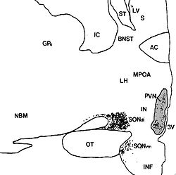
 Human paraventricular nucleus (PVN) in this coronal section is indicated by the shaded area. Dots represent vasopressin (AVP) neurons (also seen in the supraoptic nucleus, SON). The medial surface is the 3rd ventricle (3V). Human paraventricular nucleus (PVN) in this coronal section is indicated by the shaded area. Dots represent vasopressin (AVP) neurons (also seen in the supraoptic nucleus, SON). The medial surface is the 3rd ventricle (3V). | |
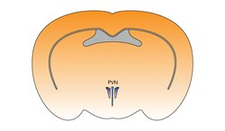
 The paraventricular hypothalamus of the mouse brain The paraventricular hypothalamus of the mouse brain | |
| Details | |
| Identifiers | |
| Latin | nucleus paraventricularis hypothalami |
| MeSH | D010286 |
| NeuroNames | 387 |
| NeuroLex ID | birnlex_1407 |
| TA98 | A14.1.08.909 |
| TA2 | 5722 |
| FMA | 62320 |
| Anatomical terms of neuroanatomy[edit on Wikidata] | |
The paraventricular nucleus of hypothalamus (PVN, PVA, or PVH) is a nucleus in the hypothalamus, that lies next to the third ventricle. Many of its neurons project to the posterior pituitary where they secrete oxytocin, and a smaller amount of vasopressin. Other secretions are corticotropin-releasing hormone (CRH) and thyrotropin-releasing hormone (TRH). CRH and TRH are secreted into the hypophyseal portal system, and target different neurons in the anterior pituitary. Dysfunctions of the PVN can cause hypersomnia in mice. In humans, the dysfunction of the PVN and the other nuclei around it can lead to drowsiness for up to 20 hours per day. The PVN is thought to mediate many diverse functions through different hormones, including osmoregulation, appetite, wakefulness, and the response of the body to stress.
Location
The paraventricular nucleus lies adjacent to the third ventricle. It lies within the periventricular zone and is not to be confused with the periventricular nucleus, which occupies a more medial position, beneath the third ventricle. The PVN is highly vascularised and is protected by the blood–brain barrier, although its neuroendocrine cells extend to sites (in the median eminence and in the posterior pituitary) beyond the blood–brain barrier. PVN is accounting for only about 1% of the brain volume.In the rat, the PVN consists of approximately 100,000 neurons located in a volume of about 0.5 cubic millimetre.
Neurons
The PVN contains magnocellular neurosecretory cells whose axons extend into the posterior pituitary, parvocellular neurosecretory cells that project to the median eminence, ultimately signalling to the anterior pituitary, and several populations of other cells that project to many different brain regions including parvocellular preautonomic cells that project to the brainstem and spinal cord.
Magnocellular neurosecretory neurons

The magnocellular cells in the PVN elaborate and secrete two peptide hormones: oxytocin and vasopressin.
These hormones are packaged into large vesicles, which are then transported down the unmyelinated axons of the cells and released from neurosecretory nerve terminals residing in the posterior pituitary gland.
Similar magnocellular neurons are found in the supraoptic nucleus which also secrete vasopressin and a smaller amount of oxytocin.
Parvocellular neurosecretory neurons
The axons of the parvocellular neurosecretory neurons of the PVN project to the median eminence, a neurohemal organ at the base of the brain, where their neurosecretory nerve terminals release their hormones at the primary capillary plexus of the hypophyseal portal system. The median eminence contains fiber terminals from many hypothalamic neuroendocrine neurons, secreting different neurotransmitters or neuropeptides, including vasopressin, corticotropin-releasing hormone (CRH), thyrotropin-releasing hormone (TRH), gonadotropin-releasing hormone (GnRH), growth hormone-releasing hormone (GHRH), dopamine (DA) and somatostatin (growth hormone release inhibiting hormone, GIH) into blood vessels in the hypophyseal portal system. The blood vessels carry the peptides to the anterior pituitary gland, where they regulate the secretion of hormones into the systemic circulation. The parvocellular neurosecretory cells include those that make:
- Corticotropin-releasing hormone (CRH), which regulates ACTH secretion from the anterior pituitary gland
- Vasopressin, which also regulates ACTH secretion (vasopressin and CRH act synergistically to stimulate ACTH secretion)
- Thyrotropin-releasing hormone (TRH), which regulates TSH and prolactin secretion
Centrally-projecting neurons
As well as neuroendocrine neurons, the PVN contains interneurons and populations of neurons that project centrally (i.e., to other brain regions). The centrally-projecting neurons include
- Parvocellular oxytocin cells, which project mainly to the brainstem and spinal cord. These neurons are thought to have a role in gastric reflexes and penile erection,
- Parvocellular vasopressin cells, which project to many points in the hypothalamus and limbic system, as well as to the brainstem and spinal cord (these are involved in blood pressure and temperature regulation), and brown fat thermogenesis.
- Parvocellular CRH neurons, which are thought to be involved in stress-related behaviors.
Afferent inputs
The PVN receives afferent inputs from many brain regions and different parts of the body, by hormonal control.
Among these, inputs from neurons in structures adjacent to the anterior wall of the third ventricle (the "AV3V region") carry information about the electrolyte composition of the blood, and about circulating concentrations of such hormones as angiotensin and relaxin, to regulate the magnocellular neurons.
Inputs from the brainstem (the nucleus of the solitary tract) and the ventrolateral medulla carry information from the heart and stomach. Inputs from the hippocampus to the CRH neurones are important regulators of stress responses.
Inputs from neuropeptide Y-containing neurons in the arcuate nucleus coordinate metabolic regulation (via TRH secretion) with regulation of energy intake. Specifically, the projections from the arcuate nucleus seem to exert their effect on appetite via MC4R-expressing oxytocinergic cells of the PVN.
Inputs from suprachiasmatic nucleus about levels of lighting (circadian rhythms).
Inputs from glucose sensors within the brain stimulate release of vasopressin and corticotropin-releasing hormone from parvocellular neurosecretory cells.
References
- Ferguson AV, Latchford KJ, Samson WK (June 2008). "The paraventricular nucleus of the hypothalamus - a potential target for integrative treatment of autonomic dysfunction". Expert Opinion on Therapeutic Targets. 12 (6): 717–27. doi:10.1517/14728222.12.6.717. PMC 2682920. PMID 18479218.
- Chen, Chang-Rui; Zhong, Yu-Heng; Jiang, Shan; Xu, Wei; Xiao, Lei; Wang, Zan; Qu, Wei-Min; Huang, Zhi-Li (2021-11-17). Elmquist, Joel K; Wong, Ma-Li; Lazarus, Michael (eds.). "Dysfunctions of the paraventricular hypothalamic nucleus induce hypersomnia in mice". eLife. 10: e69909. doi:10.7554/eLife.69909. ISSN 2050-084X. PMC 8631797. PMID 34787078.
- Wang, Zan; Zhong, Yu-Heng; Jiang, Shan; Qu, Wei-Min; Huang, Zhi-Li; Chen, Chang-Rui (2022-03-14). "Case Report: Dysfunction of the Paraventricular Hypothalamic Nucleus Area Induces Hypersomnia in Patients". Frontiers in Neuroscience. 16. doi:10.3389/fnins.2022.830474. ISSN 1662-453X. PMC 8964012. PMID 35360167.
- ^ Fox SI (2011). Human Physiology (Twelfth ed.). McGraw Hill. p. 665.
- Chen, Chang-Rui; Zhong, Yu-Heng; Jiang, Shan; Xu, Wei; Xiao, Lei; Wang, Zan; Qu, Wei-Min; Huang, Zhi-Li (2021-11-17). Elmquist, Joel K; Wong, Ma-Li; Lazarus, Michael (eds.). "Dysfunctions of the paraventricular hypothalamic nucleus induce hypersomnia in mice". eLife. 10: e69909. doi:10.7554/eLife.69909. ISSN 2050-084X. PMC 8631797. PMID 34787078.
- Grassi, D.; Marraudino, M.; Garcia-Segura, L. M.; Panzica, G. C. (2022-04-01). "The hypothalamic paraventricular nucleus as a central hub for the estrogenic modulation of neuroendocrine function and behavior". Frontiers in Neuroendocrinology. 65: 100974. doi:10.1016/j.yfrne.2021.100974. ISSN 0091-3022. PMID 34995643.
- Giuliano F, Allard J (August 2001). "Dopamine and sexual function". International Journal of Impotence Research. 13 Suppl 3: S18-28. doi:10.1038/sj.ijir.3900719. PMID 11477488.
- Argiolas A, Melis MR (May 2005). "Central control of penile erection: role of the paraventricular nucleus of the hypothalamus". Progress in Neurobiology. 76 (1): 1–21. doi:10.1016/j.pneurobio.2005.06.002. PMID 16043278. S2CID 24929538.
- Russell JA, Blackburn RE, Leng G (June 1988). "The role of the AV3V region in the control of magnocellular oxytocin neurons". Brain Research Bulletin. 20 (6): 803–10. doi:10.1016/0361-9230(88)90095-0. PMID 3044525. S2CID 4762486.
- Beck B (July 2006). "Neuropeptide Y in normal eating and in genetic and dietary-induced obesity". Philosophical Transactions of the Royal Society of London. Series B, Biological Sciences. 361 (1471): 1159–85. doi:10.1098/rstb.2006.1855. PMC 1642692. PMID 16874931.
- Konturek PC, Konturek JW, Cześnikiewicz-Guzik M, Brzozowski T, Sito E, Konturek SJ (December 2005). "Neuro-hormonal control of food intake: basic mechanisms and clinical implications" (PDF). Journal of Physiology and Pharmacology. 56 Suppl 6: 5–25. PMID 16340035.
- Nillni EA (April 2010). "Regulation of the hypothalamic thyrotropin releasing hormone (TRH) neuron by neuronal and peripheral inputs". Frontiers in Neuroendocrinology. 31 (2): 134–56. doi:10.1016/j.yfrne.2010.01.001. PMC 2849853. PMID 20074584.
- Qin C, Li J, Tang K (September 2018). "The Paraventricular Nucleus of the Hypothalamus: Development, Function, and Human Diseases". Endocrinology. 159 (9): 3458–3472. doi:10.1210/en.2018-00453. PMID 30052854.
Further reading
- Hall JE, Guyton AC (20 May 2015). Guyton and Hall Textbook of Medical Physiology. US Elsevier Health Bookshop, Mosby, Saunders, Netter & More. ISBN 9781455770052.